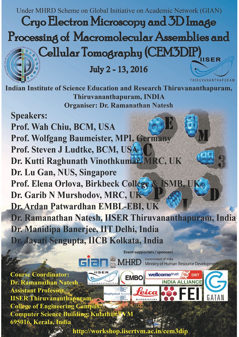
The 1st Cryo Electron Microscopy and 3D Image Processing of Macromolecular Assemblies and Cellular Tomography (CEM3DIP)
Under MHRD Scheme on Global Initiative of Academic Networks (GIAN)
|
|
The 1st Cryo Electron Microscopy and 3D Image Processing of Macromolecular Assemblies and Cellular Tomography (CEM3DIP)
Under MHRD Scheme on Global Initiative of Academic Networks (GIAN)
|
|
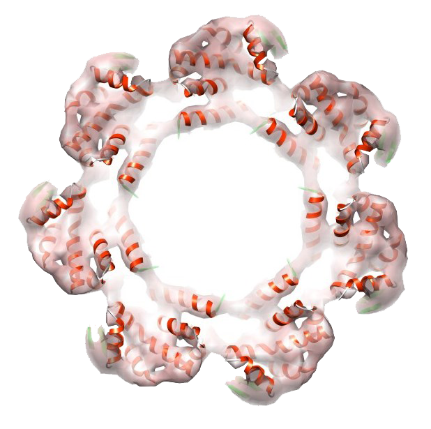 Proteins are the macromolecular machines that carry out functions of LIFE. Proteins are made of polymer of amino acids, which fold in to three-dimensional structure. Visualising and understanding the structure will help us in understanding the structure-function relationship. This will help scientists to work on medically important proteins with clear emphasis on using this knowledge to develop novel therapeutic targets/drugs. Structural Biology using Single Particle Cryo Electron Microscopy (SP CryoEM) and cellular tomography has become a major tool for studying macromolecular assemblies and visualizing unprecedented details of cellular components, respectively. Solving in-situ proteins structure is fast approaching feasiblity, with advancement in CryoEM methods. However these cutting edge CryoEM methods for structural biology are only a couple of years old in India, and there are only very few PI’s currently working in SP CryoEM field in India. This course coordinator being one of those few people who received a training in CryoEM methods as a Wellcome Trust Fellow, in Birkbeck College, London, UK and returned back to India. This course will deal with Single Particle Cryo Electron Microscopy of biological macromolecular assemblies and Cryo Electron Tomography of Cellular structures. Key words for this course would be ‘Macromolecular Assemblies, Structural Biology / Macromolecular structure by Cryo Electron Microscopy(CryoEM), Single Particle CryoEM, Cryo Electron Tomography, Multidisciplinary methods, Cryo EM as Hybrid methods in combination with Protein Crystallography’
This is a special course, which will be taught probably for the first time in India. This course will be of interest to molecular biologists, cell biologists, microbiologists, structural biologists (crystallographers), theoretical/computational biologists and in general many other area biologists and non biologists including computational scientists who are working in the field of image processing, and will be very useful at both teachers and students level.
Download the Final Time Table & Venue
Lecture notes and videos on demand of lectures L1 to L23 can be downloaded/streamed here
(Lecture videos will be uploaded soon).
|
|
|
|
|

|
|
|
|